Quick Answer: MRI machines use powerful magnets and radio waves to make detailed images of the inside of the body. They work by aligning hydrogen protons in tissues, exciting them with radiofrequency pulses, and then capturing the signals they release to form pictures with remarkable clarity—without using harmful radiation.
The Magnetic Canvas
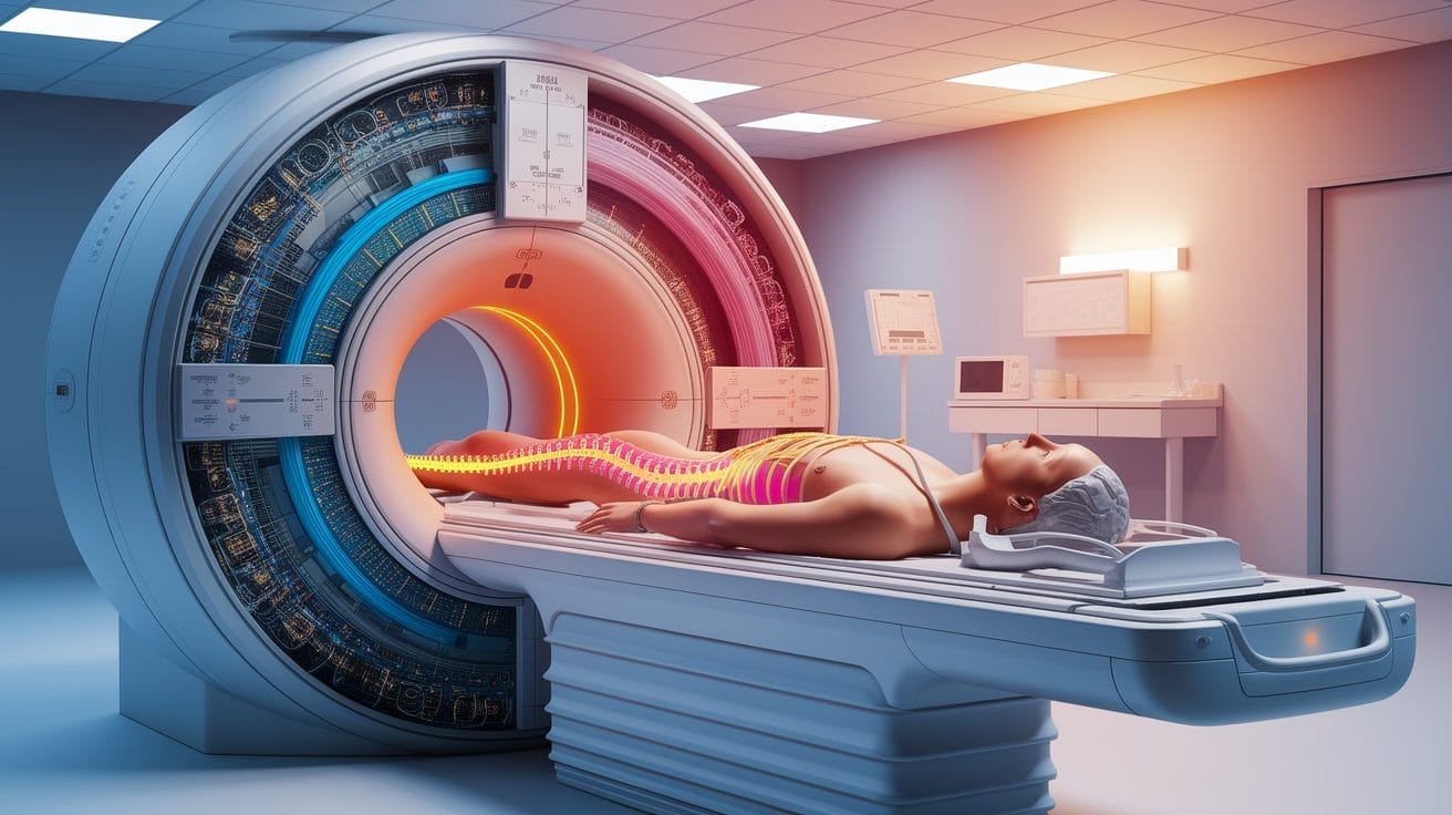
Every MRI scan begins with an incredibly strong magnetic field—often tens of thousands of times stronger than Earth’s. This magnetic field sets the stage on which the “painting” of the body’s internal landscape will be created. Why hydrogen? Because the body is mostly water, and water molecules contain hydrogen atoms, which have a single proton in their nucleus. These protons act like microscopic bar magnets.

The MRI’s superconducting magnet generates what’s called the “main field,” measured in units called Tesla. Typical clinical MRI machines operate at 1.5 or 3 Tesla strength, but experimental systems can go higher. This main magnetic field creates a uniform environment in which the protons can be precisely controlled.
Aligning Protons: Magnet Meets Matter
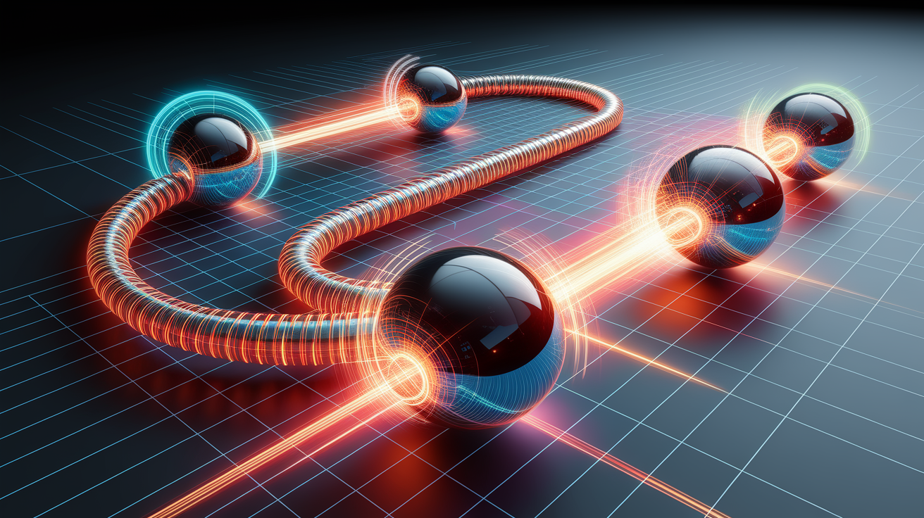
Under normal conditions, proton “mini-magnets” point in random directions. When the patient enters the MRI scanner, the magnetic field causes many of these protons to align either with or against the field. More align with it than against, creating a net magnetic effect along the field direction.

Next comes the role of radiofrequency (RF) pulses. These bursts of energy from the MRI’s RF coils are tuned to the natural resonant frequency of hydrogen atoms. When applied, they tip the aligned protons out of their equilibrium position. As soon as the RF pulse stops, the protons relax back—either through T1 relaxation (recovery along the field direction) or T2 relaxation (loss of phase coherence within the transverse plane). These different relaxation behaviors lend tissues their contrast on an MRI image.
- T1-weighted images highlight fat and provide good anatomical detail.
- T2-weighted images make fluids bright, helping detect swelling or inflammation.
Spatial Encoding: Pinpointing Signal Sources
Of course, just knowing that protons are producing signals isn’t enough—we need to know where in the body they’re coming from. This is where spatial encoding comes in. MRI machines use gradient coils to slightly vary the magnetic field strength along different axes. This makes the resonant frequency of protons depend on their position.
- Slice selection: A magnetic gradient is applied along one dimension, and the RF pulse is tuned to excite only protons in a specific slice.
- Frequency encoding: Another gradient changes proton frequencies along one axis during signal collection—allowing location mapping in that direction.
- Phase encoding: A gradient is briefly applied along the remaining axis, subtly shifting phase relationships between proton spins to encode spatial data.
By carefully sequencing these gradients and RF pulses, the scanner builds a complete map of where each signal originated, whether it’s for 2D or 3D imaging.
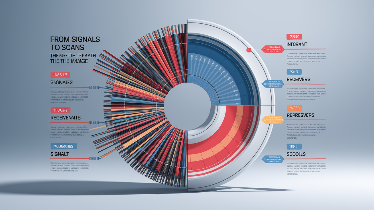
From Signals to Scans: The Math Behind the Image
The raw data picked up by the MRI’s receiver coils is not an image—it’s a complex set of signals stored in what’s called “k-space.” To turn this into the familiar cross-sectional views, the system uses mathematical tools such as the Fourier transform. This process converts variations in frequency and phase into spatial information that the computer can display as pixels or voxels in an image.
Additional image processing algorithms can sharpen image quality, reduce noise, and optimize contrast. Adjusting scan parameters like repetition time, echo time, and flip angle can further enhance detail for specific tissues or pathologies. The end result is a high-resolution map that allows clinicians to identify structures and abnormalities with remarkable precision.
Lights Out: Bringing Inner Worlds to Light
MRI’s magic lies in producing vivid images without ionizing radiation, making it ideal for repeated scans and sensitive areas like the brain and spinal cord. It’s a powerful tool for diagnosing issues from ligament tears to tumors, and for guiding treatment planning. However, safety is paramount—strong magnets can attract ferromagnetic objects and interact with certain implants such as pacemakers, so thorough screening is essential before scanning, as emphasized by the Cleveland Clinic.

Contrast agents may be used to make certain tissues or blood vessels stand out more clearly, and advanced pulse sequences can reveal physiological activity or blood flow. Continuous improvements in coil design, gradient strength, and computational power are making MRI faster, sharper, and more versatile than ever.
Wrapping Up
In essence, an MRI machine is a marvel of physics, engineering, and mathematics: it aligns tiny magnets inside our bodies, stimulates them with finely tuned radio waves, pinpoints their locations through gradient tricks, and finally reconstructs their story into images we can see. This is how powerful magnetic resonance imaging opens a safe window into the body’s hidden worlds, helping doctors answer complex questions without a single incision.